How Is TMD Diagnosed? Examination, MRI, CBCT and Finding the Real Cause
A patient can arrive with six months of jaw pain and a folder full of normal tests. Another arrives with an MRI report that sounds frightening but has only mild symptoms. Someone else has clicked for ten years and only became painful last week. A good TMD consultation is less about collecting abnormalities and more about building a coherent timeline.
The history comes before the hardware
The first questions are often the most powerful. Did the problem start suddenly or gradually? Was there trauma, dental treatment, prolonged mouth opening, illness, a change in sleep, intense work or another major event? Did clicking start before the pain? Did the click stop when locking began? Did the bite change before or after the symptoms?
Then comes provocation: chewing, yawning, hard food, talking, waking, long meetings, driving, exercise, stress, clenching or wide opening. What relieves it—rest, heat, movement, medication, massage, an appliance or avoiding hard foods? Headache, ear symptoms, neck pain, dizziness, sleep, widespread pain, prior injury, arthritis and autoimmune disease can all alter the differential diagnosis.
Movement tells a story
The clinician observes not just how far the jaw opens but how it opens. Does the mandible deviate to one side and stay there, or does it move away and return? Is the limitation hard and mechanical or soft and pain-limited? What happens with gentle assisted opening? Are lateral and protrusive movements symmetrical?
A millimetre measurement is useful, but movement quality often gives more context than the number alone.
Diagnosis is not a hunt for tender points
If enough pressure is applied, healthy people can be made tender. Modern TMD examination therefore pays particular attention to whether palpation or movement reproduces the patient’s familiar pain. Familiar pain in the chewing muscles supports a muscular diagnosis; familiar pain at the joint with appropriate palpation or function supports arthralgia. Many patients have a mixed pattern.
When imaging is not necessary
Not every TMD requires MRI or CBCT. If the history and examination fit a common myofascial pain disorder, there are no red flags and no important mechanical findings, imaging may not change initial treatment. An expensive image that does not change the plan is not automatically better care.
When CBCT helps
CBCT is excellent for bony detail. It may be useful when the clinician needs to evaluate suspected degenerative changes, significant asymmetry, trauma, ankylosis, developmental abnormality, surgical anatomy or another question where bone information will change management. It can show flattening, erosion, sclerosis, osteophytes and fractures.
When MRI helps
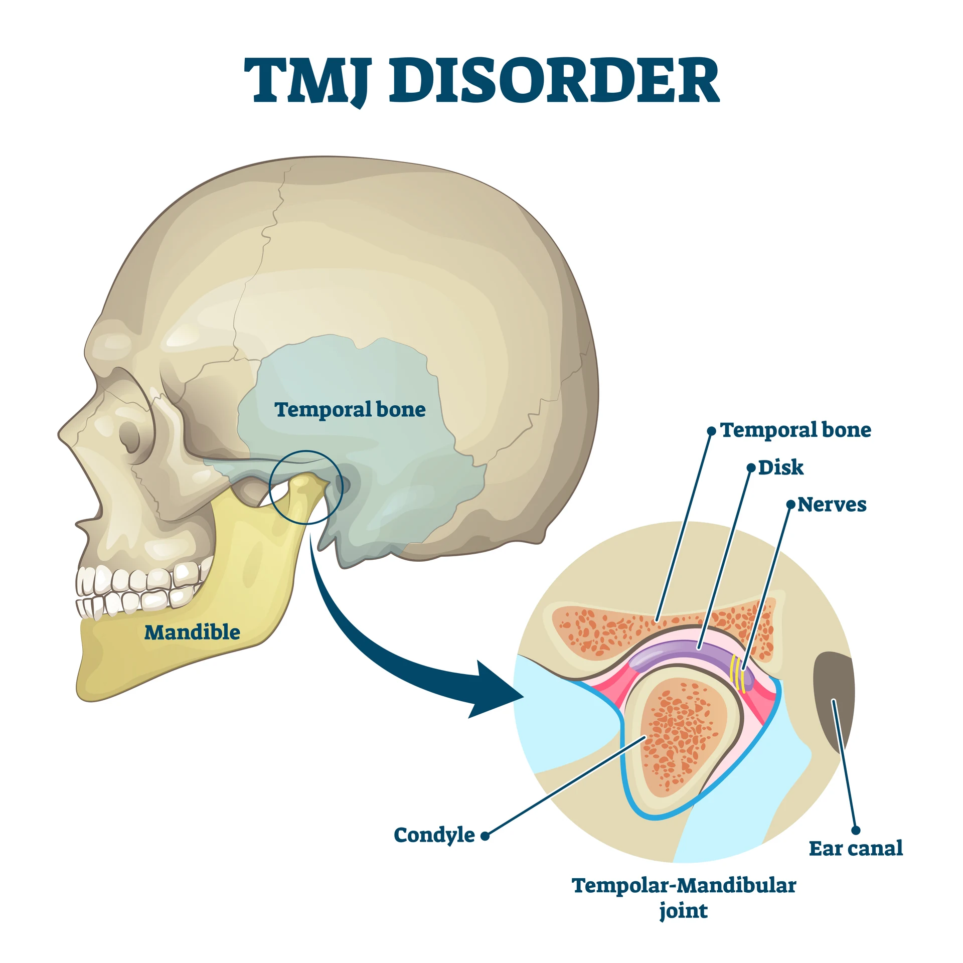
MRI is the principal clinical imaging modality for the TMJ disc and other soft tissues. It can assess disc position, joint effusion and soft-tissue relationships in closed- and open-mouth positions depending on the protocol. Persistent locking and a clinically important internal-derangement question are very different indications from a painless click.
What a scan cannot tell you
An MRI can show a displaced disc; it cannot prove by itself that the disc is the main reason for the pain. A CBCT can show osteophytes; it cannot tell the clinician how much pain a patient experiences. Structural abnormalities may exist without major symptoms, while severe pain can occur without dramatic structural change. The scan is evidence—not the verdict.
Where digital bite analysis fits
T-Scan and similar systems can document occlusal contact timing and relative force distribution. That can be useful when a specific restorative or bite question is part of the case. But digital occlusal analysis does not establish myalgia, arthralgia, disc displacement or degenerative joint disease. A sophisticated graph cannot compensate for a weak diagnosis.
The conditions a TMD clinician must not miss
Dental disease remains one of the most important alternatives: pulp disease, cracked teeth, periodontal disease and pericoronitis can refer pain into the jaw and face. Migraine and other headache disorders can coexist with TMD. ENT disease, neuropathic pain such as trigeminal neuralgia, inflammatory arthritis, infection, fracture, salivary disease and tumors can also mimic or complicate the picture.
Red flags change the tempo. Progressive unexplained restriction, persistent swelling, fever, a mass, sensory or motor changes, significant trauma, unexplained weight loss or persistent non-mechanical night pain should prompt additional investigation or referral.
The diagnostic sentence
At the end of an assessment, “You have TMJ” is not enough. A useful diagnosis sounds more like: “Your examination is most consistent with pain from the chewing muscles, with daytime jaw bracing and neck pain contributing,” or “Your history suggests an intra-articular problem with restricted opening, and MRI may alter the next step.” A good diagnosis is a working model that predicts what should help and tells us what to reassess if improvement does not occur.
Treatment should follow the diagnosis—not the other way around.
The complete “Finding the Real Problem” chapter in Understanding Your TMJ turns this diagnostic process into a patient-friendly roadmap: what to tell your clinician, what the examination is trying to reproduce, when MRI or CBCT is useful, and what other diagnoses must be considered. If you have already had multiple scans or treatments but still cannot answer “what exactly is my diagnosis?”, this may be the most important chapter for you to read.